
The ability to wiggle your toes with a broken foot can often be an intriguing indicator of the injury’s nature. While toe movement may still be possible, it is not necessarily an indicator of the fracture’s severity. This phenomenon is largely due to the complex interplay of nerves, muscles, and bones within the foot. Understanding the anatomy of the foot and the types of fractures that can occur is essential for appreciating why toe mobility might persist even when a significant injury is present. What implications does this have for diagnosing and treating foot fractures?
Anatomy of the Foot
The anatomy of the foot comprises 26 bones, 33 joints, and more than 100 muscles, tendons, and ligaments, each contributing to its complex structure and function. This intricate assembly provides the necessary support, balance, and mobility critical for ambulation and weight distribution. The bones of the foot are categorized into three groups: tarsal bones, metatarsal bones, and phalanges. The tarsal bones form the hindfoot and midfoot, while the metatarsals and phalanges constitute the forefoot.
Foot arches play a pivotal role in absorbing shock and maintaining balance. The medial and lateral longitudinal arches, along with the transverse arch, distribute body weight across the foot. These arches are maintained by the ligament connections, particularly the plantar fascia and the spring ligament, which offer structural integrity and elasticity.
Ligament connections, such as the deltoid ligament and the calcaneonavicular ligament, facilitate joint stability and motion control. The interplay between these ligaments and the surrounding muscles, including the intrinsic and extrinsic muscle groups, allows for complex movements and adjustments necessary for varied activities. Understanding this anatomical complexity is essential for diagnosing and managing foot injuries effectively.
Types of Foot Fractures
Understanding the various types of foot fractures is critical for effective treatment and recovery. Stress fractures, resulting from repetitive force, differ notably from compound and simple fractures, which involve varying degrees of bone displacement and skin penetration. Additionally, diagnosing hairline fractures requires advanced imaging techniques due to their subtle presentation on initial radiographs.
Stress Fractures Explained
Stress fractures, commonly seen in athletes and military recruits, are small cracks in the bone caused by repetitive force or overuse. These injuries often result from cumulative bone stress that the skeletal system cannot adequately repair, leading to a fatigue injury. Basically, the repetitive microtrauma surpasses the bone’s intrinsic ability to remodel and strengthen, culminating in a stress fracture.
Clinically, stress fractures manifest as localized pain that intensifies with weight-bearing activities and diminishes with rest. Diagnostic imaging, particularly MRI and bone scintigraphy, can detect these fractures more sensitively than plain radiographs, especially in the early stages. The most frequently affected bones in the foot include the second and third metatarsals, the navicular, and the calcaneus.
Risk factors for stress fractures encompass intrinsic elements such as bone density and biomechanics, as well as extrinsic factors like training intensity and footwear. Management typically involves a period of rest and activity modification, allowing for bone healing. In some cases, immobilization or the use of a pneumatic walking boot may be necessary to alleviate stress on the affected bone. Early diagnosis and intervention are vital to prevent progression and ensure optimal recovery.
Compound Vs. Simple Fractures
Compound and simple fractures represent two primary classifications of foot fractures, distinguished by whether the bone pierces the skin and the complexity of the injury. A compound fracture, also known as an open fracture, occurs when the broken bone disrupts the skin, leading to a higher risk of infection and requiring more complex medical management. This type of fracture is often associated with significant trauma, necessitating surgical intervention to realign the bone fragments and guarantee appropriate healing.
In contrast, a simple fracture, or closed fracture, involves a break in the bone without an associated open wound. This type of fracture can often be managed with immobilization techniques such as casting or splinting, provided the bone fragments remain well-aligned. Factors such as bone density play a substantial role in influencing the likelihood and severity of foot fractures. Reduced bone density, as seen in conditions like osteoporosis, increases the susceptibility to both compound and simple fractures.
The fracture classification also considers the specific location and pattern of the break. Understanding these distinctions is vital for developing an effective treatment plan and optimizing patient outcomes. Accurate diagnosis and timely intervention are paramount in preventing complications and ensuring the best possible recovery.
Diagnosing Hairline Fractures
Hairline fractures, also known as stress fractures, are minute cracks in the bone that often require advanced imaging techniques for accurate diagnosis. These fractures are typically the result of repetitive stress or overuse, often seen in athletes or individuals with compromised bone density. Standard X-rays may not always detect these microfractures due to their diminutive size and the subtlety of the associated bone changes.
Microfracture detection often necessitates the use of more sensitive imaging modalities such as Magnetic Resonance Imaging (MRI) or bone scans. MRI provides high-resolution images of both bone and surrounding soft tissues, making it particularly effective in identifying stress fractures. Bone scans, which involve the injection of a small amount of radioactive material, can reveal areas of increased bone metabolism indicative of a fracture.
Bone density plays an important role in the susceptibility to hairline fractures. Individuals with lower bone density, such as those with osteoporosis, are at a heightened risk. Dual-energy X-ray absorptiometry (DEXA) scans are commonly used to assess bone density, aiding in the evaluation of fracture risk and the formulation of preventive strategies. Accurate and timely diagnosis is essential for effective management and to prevent progression to more severe fractures.
Symptoms of a Broken Foot
Patients with a broken foot often present with symptoms such as acute localized pain, swelling, and difficulty bearing weight on the affected limb. Swelling identification is crucial for clinicians, as it can indicate the severity and specific location of the fracture. Swelling typically manifests soon after the injury and may continue to increase over the subsequent hours. The presence of edema can obscure anatomical landmarks, complicating the diagnosis and necessitating further imaging studies such as X-rays or MRIs.
Pain localization is another essential aspect of evaluating a broken foot. The pain is typically sharp and concentrated at the site of the fracture. Patients may report exacerbated pain upon palpation of the affected area or when attempting to move the foot. This pain often radiates and can be associated with bruising or a visible deformity, depending on the fracture’s nature and severity.
Additionally, patients frequently experience functional impairment, including an inability to bear weight on the injured foot. This symptom is often accompanied by an altered gait or complete inability to ambulate. These clinical manifestations require prompt evaluation and intervention to prevent further complications and ensure optimal recovery.
Toe Movement and Fractures
Toe movement can provide valuable diagnostic information in the assessment of foot fractures. The presence of toe flexibility suggests that certain structures within the foot remain intact despite the fracture. Clinically, the ability to wiggle the toes may indicate that the tendons, ligaments, and muscles responsible for toe movement are not severely compromised. However, the degree of toe movement does not necessarily correlate with the severity of the fracture.
Fracture pain often becomes pronounced upon attempting to move the toes due to the transmission of mechanical stress to the fracture site. This can exacerbate discomfort and highlight the need for further imaging studies, such as X-rays or MRI, to ascertain the extent of bone damage. Clinicians should note that while some patients with foot fractures retain a surprising amount of toe flexibility, others may experience limited movement due to significant swelling, soft tissue injury, or pain inhibition.
Differentiating between types of fractures—such as non-displaced versus displaced fractures—can also inform the expected range of toe movement. Non-displaced fractures, where bone fragments remain aligned, may present with more preserved toe flexibility compared to displaced fractures. Ultimately, a thorough clinical evaluation, including the assessment of toe movement, is essential for accurate diagnosis and effective management of foot fractures.

Nerve and Muscle Function
Proper nerve and muscle function is critical in evaluating the overall impact of foot fractures on mobility and recovery. A thorough understanding of nerve pathways and muscle coordination is essential for clinicians to assess the extent of impairment and devise effective treatment plans. The foot’s intricate network of nerves, including the tibial and peroneal nerves, plays a pivotal role in transmitting signals between the brain and the muscles responsible for toe movement.
When a fracture occurs, these nerve pathways can be compromised, leading to altered sensory and motor functions. Disruption in nerve conduction may result in diminished proprioception, affecting the coordinated movement of the toes. Clinically, this can present as weakness, numbness, or an inability to perform fine motor tasks with the toes.
Muscle coordination, governed by the interplay between neural signals and muscular response, is also critical. The intrinsic and extrinsic muscles of the foot must work in harmony to achieve precise toe movements. Any impairment in this coordination due to nerve damage can hinder the rehabilitation process. As such, a thorough neurological examination is indispensable in the clinical assessment of foot fractures to ensure optimal functional recovery and prevent long-term disability.
Severity of the Injury
Evaluating the severity of a foot fracture requires a thorough examination of both the structural damage and the degree of compromised nerve and muscle function. Clinicians typically employ radiographic imaging, such as X-rays or MRIs, to ascertain the extent of the fracture. The classification of the fracture—whether it is a simple, compound, comminuted, or displaced fracture—significantly influences the treatment protocol and expected outcomes.
Pain levels are a critical indicator of severity. Higher pain levels often correlate with more extensive damage, although this is not always the case due to individual pain tolerance and variability in nerve involvement. Severe fractures can compromise blood flow and nerve function, further complicating the clinical picture.
Healing time is another key factor in evaluating the severity of the injury. Simple fractures may heal within six to eight weeks with appropriate immobilization and limited weight-bearing activities. Conversely, complex fractures, particularly those involving multiple bone fragments or misalignment, may require surgical intervention and a more extended recovery period, often exceeding three months. These cases may also necessitate physical therapy to restore function and strength. Understanding the severity helps in setting realistic expectations for recovery and informing the patient about potential complications.
Stress Fractures Explained
Stress fractures, characterized by small cracks or severe bruising within a bone, often result from repetitive force or overuse, particularly in weight-bearing activities. These injuries are prevalent among athletes, military recruits, and individuals with occupations requiring continuous physical exertion. Clinically, stress fractures are most commonly observed in the metatarsal bones of the foot, tibia, and femoral neck.
The pathophysiology of stress fractures involves an imbalance between bone resorption and formation. Repetitive stress leads to micro-damage that exceeds the bone’s capacity to repair itself, thereby resulting in a fracture. Bone density plays a critical role in this process; individuals with lower bone density, such as those with osteoporosis, are at a heightened risk. Conversely, individuals with optimal bone density are better equipped to withstand repetitive stress, thereby reducing the likelihood of fracture.
Diagnostic imaging, including X-rays, MRI, and bone scans, is essential for confirming the presence of a stress fracture. Evidence-based treatment protocols typically involve activity modification, immobilization, and in certain cases, pharmacologic interventions to enhance bone healing. Understanding the etiology and risk factors associated with stress fractures is paramount for effective prevention and management strategies in clinical practice.
Diagnosing a Broken Foot
Accurate diagnosis of a broken foot necessitates a thorough clinical evaluation alongside advanced imaging techniques to determine the extent and specific location of the fracture. The initial medical consultation involves a detailed patient history and a physical examination to assess symptoms such as pain, swelling, bruising, and functional impairments. The clinician may palpate the foot to identify areas of tenderness, deformity, or crepitus that indicate a potential fracture.
Subsequently, imaging techniques are essential to confirm the diagnosis and to provide a precise assessment of the fracture characteristics. Standard radiographs (X-rays) are typically the first-line imaging modality and can effectively reveal most fractures. However, in cases where the fracture is not clearly visible on X-rays, advanced imaging techniques such as computed tomography (CT) scans or magnetic resonance imaging (MRI) may be employed. CT scans offer detailed cross-sectional images that are particularly useful for evaluating complex fractures, while MRIs provide superior soft tissue contrast, aiding in the detection of stress fractures and associated injuries.
A timely and accurate diagnosis through these clinical and imaging methods is critical for guiding appropriate management and facilitating ideal recovery. Further steps and considerations for treatment will be discussed in the subsequent section.

Treatment Options
Effective management of a broken foot encompasses several treatment modalities, each tailored to the severity and type of fracture. Primary options include immobilization and rest to facilitate natural bone healing, surgical intervention for complex or displaced fractures, and physical therapy to restore function and strength post-healing. Evidence-based guidelines emphasize a multidisciplinary approach to optimize recovery outcomes.
Immobilization and Rest
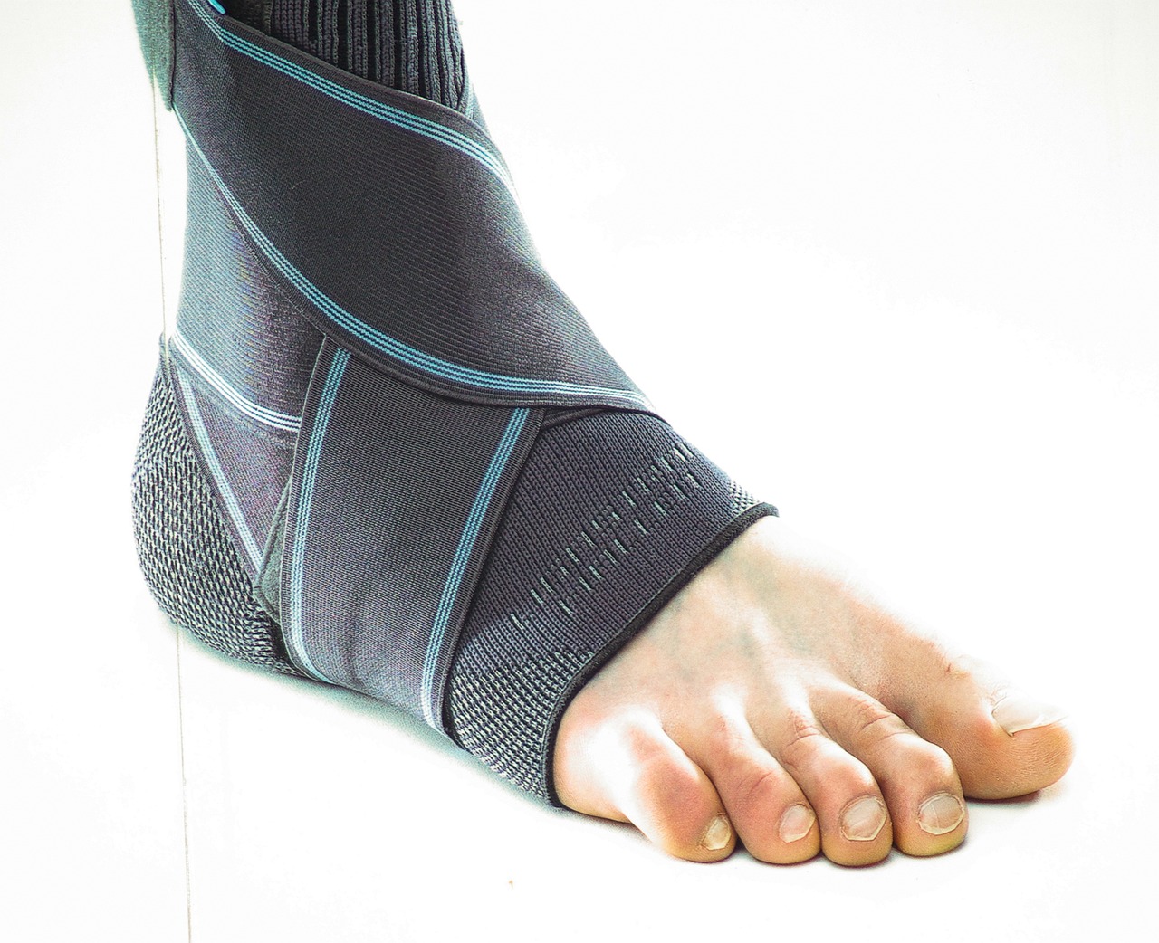
Immobilization and rest are fundamental treatment approaches for managing a broken foot, aimed at stabilizing the fracture and promoting ideal healing. Effective immobilization typically involves the use of a cast or splint to restrict movement, thereby reducing the risk of exacerbating the injury. The specific type of immobilization device is determined based on the location and severity of the fracture.
Crutch usage is often recommended to enforce weight bearing restrictions, which are vital in mitigating stress on the fractured site. Non-weight bearing (NWB) status is generally advised initially to prevent displacement of the fracture fragments and to allow the bone to heal correctly. As healing progresses, partial weight bearing (PWB) may be introduced under strict clinical supervision, contingent upon radiographic evidence of bone union and patient-reported symptom amelioration.
Adjunctive measures such as elevation and cryotherapy can further aid in reducing edema and alleviating pain. Compliance with these protocols is essential to avoid complications such as non-union or malunion. Immobilization and rest, combined with appropriate weight bearing restrictions and crutch usage, form the cornerstone of conservative management for a broken foot, optimizing the conditions for natural bone regeneration and functional recovery.
Surgical Intervention
In cases where conservative management fails to achieve satisfactory outcomes or when the fracture is particularly severe, surgical intervention becomes a necessary consideration. The primary objective of surgical treatment is to achieve ideal anatomical alignment and stabilization, thereby facilitating proper healing and functional restoration of the foot.
Surgical techniques vary depending on the type and location of the fracture. Open reduction and internal fixation (ORIF) is commonly employed for displaced fractures, involving the realignment of bone fragments and the use of internal hardware to maintain stabilization. Implant choice is critical in ORIF, with options including screws, plates, and intramedullary nails, each selected based on the specific fracture characteristics and patient needs.
In more complex fractures, such as comminuted or intra-articular fractures, external fixation may be utilized. This involves the placement of pins or screws into the bone, connected externally by a stabilizing frame. External fixation provides the benefit of minimizing soft tissue damage while maintaining alignment.
Post-operative care is essential for ideal recovery, with weight-bearing restrictions and immobilization often required to protect the surgical site. Regular follow-up and imaging are necessary to monitor healing and to identify any complications early.
Physical Therapy
Physical therapy plays a crucial role in the rehabilitation process following a broken foot, focusing on restoring strength, flexibility, and functional mobility. Once the fracture has sufficiently healed, a tailored physical therapy regimen will include a series of rehabilitation exercises designed to enhance muscle strength and joint flexibility. These exercises are essential for preventing atrophy and ensuring that the foot regains its full range of motion.
Initially, passive range-of-motion exercises may be employed, gradually evolving to active exercises as the patient’s condition improves. Strengthening exercises, such as resistance band work and proprioceptive training, are incorporated to bolster the intrinsic and extrinsic muscles of the foot. Additionally, weight-bearing activities are progressively introduced to enhance bone density and support structural integrity.
Mobility aids, such as crutches or walking boots, are often used to facilitate safe ambulation during the early stages of recovery. As the patient progresses, these aids are gradually phased out to encourage independent mobility. Evidence-based practice highlights the importance of patient-specific protocols, continuous assessment, and adjustment of the rehabilitation plan to optimize outcomes. Consistent adherence to the prescribed physical therapy regimen significantly enhances the likelihood of a full recovery, enabling patients to resume their normal activities.
Recovery Process
The recovery process for a broken foot involves a structured regimen of rest, immobilization, and gradual reintroduction of movement to facilitate best healing. Initially, the primary focus is immobilization, often achieved through the use of a cast or a splint, to ensure proper alignment and stabilization of the fractured bones. Pain management is essential during this phase, typically involving the administration of analgesics and anti-inflammatory medications to mitigate discomfort and inflammation.
The recovery timeline varies depending on the severity and location of the fracture, as well as the patient’s overall health. Minor fractures may require immobilization for approximately 6-8 weeks, whereas more complex fractures could necessitate a longer duration. Following immobilization, physical therapy becomes integral to restore function and strength, involving exercises designed to enhance range of motion and muscle conditioning.
Gradual weight-bearing activities are introduced based on clinical assessments and radiographic evidence of bone healing. Adherence to a physician-supervised rehabilitation protocol is vital to avoid complications such as non-union or malunion of the fracture. Throughout the process, periodic evaluations and adjustments to the treatment plan ensure optimal recovery, aiming to return the patient to full functional capacity.
When to Seek Help
Recognizing the appropriate time to seek medical intervention is vital for ensuring excellent outcomes in the management of a broken foot. Timely intervention can greatly influence the healing process and prevent complications such as malunion or chronic pain. Clinical indicators necessitating a medical consultation include severe pain unrelieved by rest, ice, and elevation, visible deformity, or an inability to bear weight on the affected foot. Additionally, signs of circulatory compromise, such as pallor, cyanosis, or diminished pulses in the foot, warrant immediate evaluation.
Radiographic imaging, primarily X-rays, remains the gold standard for diagnosing fractures. Advanced imaging modalities, including MRI or CT scans, may be employed for complex or occult fractures. Early diagnosis facilitates appropriate management, which may range from conservative measures like immobilization and non-weight bearing to surgical intervention for displaced fractures.
Healthcare professionals should also monitor for signs of compartment syndrome, a potentially life-threatening condition characterized by escalating pain, paresthesia, and tense swelling. Prompt recognition and surgical decompression are imperative.
Frequently Asked Questions
Can a Broken Foot Cause Long-Term Complications in Toe Movement?
Yes, a broken foot can potentially cause long-term complications in toe movement due to nerve damage. Timely intervention and consistent physical therapy are critical to mitigate these risks and promote peak recovery and functionality.
How Does Footwear Affect Recovery From a Broken Foot?
Proper footwear types notably influence recovery speed from a broken foot. Specialized orthopedic shoes or boots provide necessary support and immobilization, reducing stress on the fracture site, thereby promoting efficient healing and minimizing the risk of complications.
Are There Exercises to Regain Toe Mobility After a Foot Fracture?
Yes, there are exercises to regain toe mobility after a foot fracture. Physical therapy protocols often include specific stretching exercises designed to enhance flexibility, strength, and range of motion in the affected toes, promoting ideal recovery.
Can Broken Foot Lead to Arthritis in the Future?
A broken foot can potentially lead to arthritis in the future due to joint inflammation and cartilage damage. These complications may cause long-term changes in joint structure and function, increasing the risk of developing post-traumatic arthritis.
What Role Does Diet Play in Healing a Broken Foot?
Diet plays a crucial role in healing a broken foot by enhancing nutrient absorption and promoting tissue repair. Increased protein intake supports collagen synthesis and bone regeneration, thereby expediting the recovery process and improving overall healing outcomes.