
Minimally Invasive Lumbar Discectomy represents a significant advancement in spine surgery, particularly for patients suffering from nerve compression due to herniated or degenerative discs in the lumbar region. Unlike traditional open surgeries, which often involve extensive tissue disruption and prolonged recovery, this innovative approach utilizes small incisions, specialized instruments, and fluoroscopic guidance to precisely target and remove the problematic disc material. The benefits include reduced recovery times, minimal scarring, and a quicker return to normal activities. However, understanding the nuanced differences between this and traditional methods, as well as the potential risks involved, is essential for those considering this surgical option.
Understanding Lumbar Discectomy
A lumbar discectomy is a surgical procedure aimed at removing a portion of a herniated or degenerative disc in the lower spine to alleviate nerve compression and associated symptoms. This procedure is typically considered when thorough treatments, such as physical therapy, medications, and epidural steroid injections, have failed to provide adequate relief. Surgical alternatives to lumbar discectomy include spinal fusion, artificial disc replacement, and laminectomy; however, discectomy remains a favorable option due to its minimally invasive nature and relatively quick recovery time.
Patient eligibility for lumbar discectomy is determined through a detailed evaluation, including a comprehensive medical history, physical examination, and imaging studies such as MRI or CT scans. Ideal candidates typically present with significant radiculopathy, motor weakness, or neurological deficits directly attributable to disc herniation. Additionally, the severity and duration of symptoms, as well as the patient’s overall health, play important roles in determining suitability for surgery.
Clinical studies have demonstrated that lumbar discectomy provides substantial symptomatic relief and improved functional outcomes for appropriately selected patients. Nonetheless, patient-specific factors must be carefully considered to maximize the procedure’s efficacy and minimize potential complications.
Causes of Herniated Discs
Herniated discs are primarily caused by degenerative changes in the spine, often exacerbated by factors such as repetitive strain, trauma, and genetic predisposition. Degenerative disc disease, a natural consequence of aging, leads to the gradual deterioration of intervertebral disc integrity. This degradation results in decreased disc height and elasticity, rendering the disc more susceptible to herniation under mechanical stress.
Lifestyle factors, including sedentary behavior, poor posture, and inadequate ergonomic practices, greatly contribute to the incidence of herniated discs. Chronic repetitive strain from occupational activities or recreational pursuits can impose excessive mechanical load on the lumbar spine, accelerating disc degeneration and predisposing individuals to disc herniation. Additionally, trauma, such as falls or vehicular accidents, can precipitate acute disc herniation by imparting sudden, intense forces on the spinal column.
Genetic predisposition plays a critical role in an individual’s susceptibility to herniated discs. Studies have demonstrated that variations in genes responsible for the structural proteins of intervertebral discs, such as collagen and proteoglycans, may influence disc resilience and propensity for degeneration. Understanding these etiological factors is paramount for developing preventive strategies and optimizing therapeutic interventions, such as minimally invasive lumbar discectomy, to mitigate the impact of herniated discs on patient health.
Symptoms of Lumbar Disc Issues
Patients with lumbar disc issues commonly present with pain localized in the lower back, which may radiate to the buttocks, thighs, and legs. Symptoms often include numbness and tingling sensations in the affected areas, indicative of nerve compression. Additionally, muscle weakness in the lower extremities may be observed, potentially affecting gait and mobility.
Common Pain Locations
Thorough evaluation is required for diagnosing lumbar disc issues commonly manifesting as pain localized in the lower back, radiating to the buttocks, thighs, and sometimes extending to the lower legs and feet. This pain often results from radicular pain, which occurs when a herniated disc compresses one or more lumbar nerve roots. Radicular pain is frequently accompanied by sciatic discomfort, characterized by a sharp, burning sensation that travels along the sciatic nerve pathway.
The origins of such pain can be traced to intervertebral disc herniation, which causes mechanical compression and chemical irritation of the affected nerve roots. Clinical evidence suggests that the L4-L5 and L5-S1 disc levels are the most commonly implicated in generating these pain patterns. The distribution of pain is contingent on the specific nerve root involved, with L5 radiculopathy causing pain that radiates to the outer thigh, calf, and dorsum of the foot, while S1 radiculopathy manifests as discomfort extending to the posterior thigh, calf, and lateral foot.
Accurate diagnosis necessitates a thorough clinical evaluation, including a detailed patient history and physical examination, supplemented by imaging modalities such as MRI. Early identification and targeted therapeutic intervention are critical to mitigating symptoms and enhancing patient outcomes.
Numbness and Tingling
Numbness and tingling in the lower extremities are hallmark symptoms of nerve root compression due to intervertebral disc herniation. These sensory disturbances are frequently attributed to mechanical pressure exerted by herniated disc material on the adjacent nerve roots, leading to disrupted signal transmission. The resultant paresthesia typically follows a dermatomal distribution pattern, correlating with the specific nerve root affected. For example, compression of the L5 nerve root often manifests as numbness and tingling extending into the dorsal aspect of the foot and big toe.
Differentiating between lumbar disc-related neuropathy and other etiologies such as peripheral neuropathy is essential for accurate diagnosis and treatment planning. Peripheral neuropathy, often caused by systemic conditions like diabetic neuropathy, presents with a more generalized and symmetrical sensory loss, typically beginning in the feet and progressing proximally. In contrast, lumbar disc herniation-induced symptoms are usually unilateral and localized.
Clinical evaluation, including a detailed patient history and physical examination, is imperative to distinguish between these conditions. Diagnostic imaging, such as MRI, can confirm nerve root compression, while nerve conduction studies may be employed to rule out peripheral neuropathy. Accurate diagnosis guides effective intervention, including minimally invasive lumbar discectomy, to alleviate nerve compression and resolve sensory disturbances.
Muscle Weakness Signs
In addition to sensory disturbances, motor deficits such as muscle weakness are a significant clinical manifestation of nerve root compression from lumbar disc herniation. Patients often present with reduced strength in specific muscle groups innervated by the affected nerve roots. This weakness can manifest as difficulty in performing routine activities, such as walking on heels or toes, climbing stairs, or standing from a seated position. These motor deficits underscore the importance of early and accurate diagnosis.
Diagnostic tests play an essential role in evaluating muscle weakness associated with lumbar disc issues. Electromyography (EMG) and nerve conduction studies (NCS) can quantify the extent of nerve impairment and localize the lesion. Magnetic Resonance Imaging (MRI) is invaluable for visualizing disc herniation and its impact on neural structures. These tests facilitate a thorough assessment, guiding therapeutic interventions.
Physical therapy is integral to the management of muscle weakness secondary to lumbar disc herniation. A tailored physical therapy regimen aims to strengthen affected muscle groups, enhance flexibility, and improve functional mobility. Techniques such as neuromuscular re-education and progressive resistance exercises are employed to mitigate deficits and optimize recovery. Consequently, physical therapy not only alleviates symptoms but also enhances overall quality of life.
Traditional Surgery Vs. Minimally Invasive
While traditional lumbar discectomy involves an extensive open surgical approach with substantial tissue disruption, minimally invasive techniques focus on reducing operative trauma and enhancing recovery outcomes. Traditional discectomy necessitates a larger incision, often leading to increased muscle and soft tissue damage, prolonged hospital stays, and extended recovery periods. In contrast, minimally invasive lumbar discectomy employs smaller incisions, specialized instruments, and often endoscopic assistance, resulting in markedly reduced muscle injury and faster postoperative recovery.
Cost comparison between the two approaches reveals a nuanced picture. Initial costs for minimally invasive procedures may be higher due to specialized equipment and training; however, overall cost-effectiveness is often achieved through shorter hospital stays and quicker return to normal activities. Studies have shown that patients undergoing minimally invasive lumbar discectomy typically experience less postoperative pain, reduced need for analgesics, and lower incidence of complications, thereby potentially decreasing long-term healthcare expenses.
Furthermore, the reduced length of hospital stay associated with minimally invasive techniques not only contributes to lower overall healthcare costs but also minimizes the risks of hospital-acquired infections and other inpatient complications. This approach aligns with current healthcare objectives aimed at enhancing patient outcomes while optimizing resource utilization.

Preparing for the Procedure
Before undergoing minimally invasive lumbar discectomy, patients must undergo a thorough preoperative evaluation to guarantee successful surgical outcomes and mitigate potential risks. This evaluation typically includes a detailed medical history, physical examination, and imaging studies such as MRI or CT scans to precisely localize the herniated disc.
A critical component of preparation is the pre op checklist, which assures that all necessary steps are taken prior to the surgery. The checklist includes confirming the patient’s medical history, reviewing any current medications, and addressing any comorbid conditions that may impact anesthesia or postoperative recovery.
Dietary restrictions are another essential aspect of preoperative preparation. Patients are generally advised to refrain from eating or drinking for a specified period before the surgery, typically 8-12 hours, to reduce the risk of aspiration during anesthesia. Additionally, specific instructions regarding the discontinuation of certain medications, particularly anticoagulants and antiplatelet agents, are provided to minimize intraoperative bleeding risks.
The Surgical Process
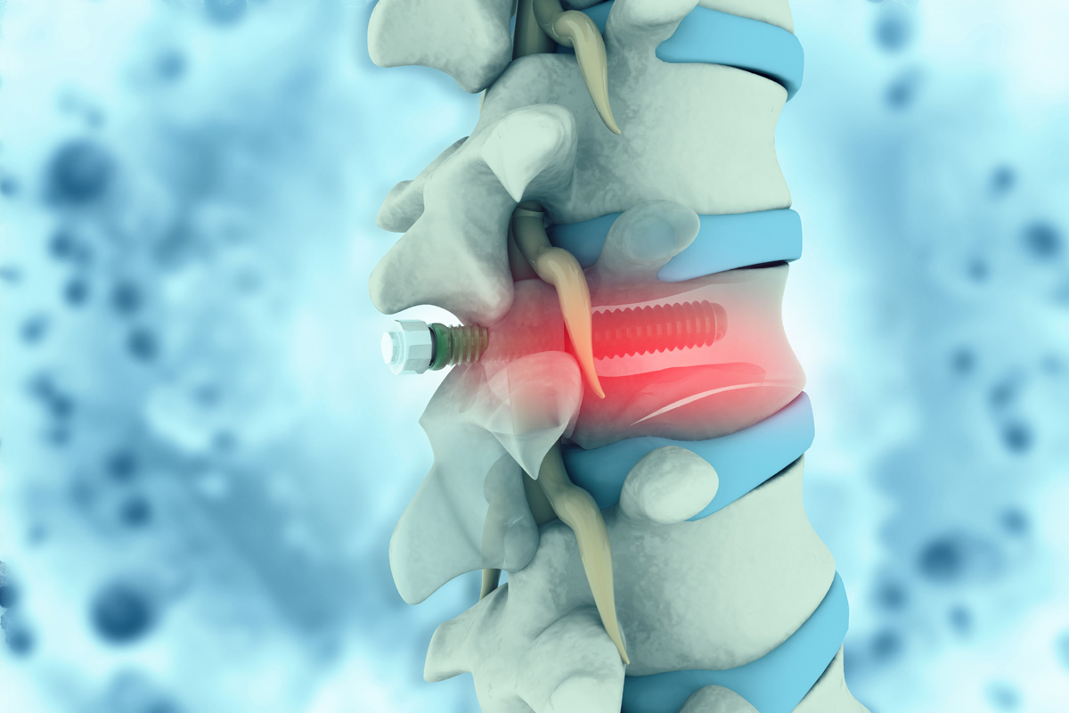
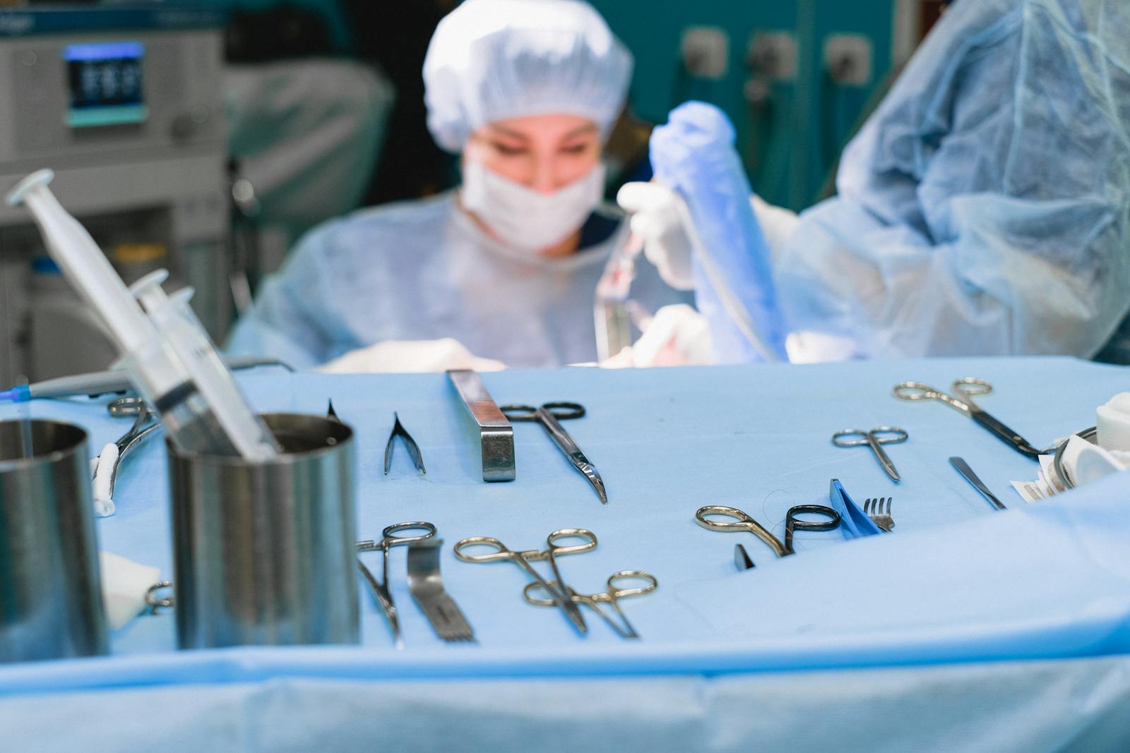
The surgical process of a minimally invasive lumbar discectomy involves making a small incision and using specialized instruments to remove the herniated disc material compressing the spinal nerves. Initially, anesthesia administration is carried out to guarantee the patient remains unconscious and pain-free throughout the procedure. General anesthesia is commonly employed, although regional anesthesia may be utilized based on the patient’s medical history and the surgeon’s discretion.
Following anesthesia, meticulous instrument sterilization is crucial to prevent postoperative infections. The surgical area is then prepared and draped in a sterile fashion. A small incision, typically less than 2 centimeters, is made in the patient’s back. Through this incision, a tubular retractor is inserted to create a pathway to the herniated disc with minimal disruption to surrounding tissues.
Under fluoroscopic guidance, specialized micro-instruments and an endoscope are used to visualize the operative field. The surgeon carefully excises the herniated disc material while preserving the integrity of the vertebral structures. The use of high-definition imaging and precision tools ensures maximal decompression of the affected nerve roots with minimal collateral damage. Once the disc material is removed, the instruments are withdrawn, and the incision is closed with sutures or surgical adhesive.
Recovery and Aftercare
Following a minimally invasive lumbar discectomy, patients typically experience a shorter recovery period and reduced postoperative pain compared to traditional open surgery. This accelerated convalescence is largely attributed to the reduced tissue disruption inherent in minimally invasive techniques. Early mobilization is encouraged, with patients often ambulating within hours post-surgery. However, a structured regimen of postoperative exercises is essential to restore function and prevent complications. These exercises, designed by physical therapists, focus on strengthening the core and lumbar musculature, enhancing spinal stability.
Dietary adjustments also play a significant role in postoperative recovery. A balanced diet rich in protein, vitamins, and minerals supports tissue healing and reduces inflammation. Increased fiber intake is recommended to mitigate constipation, a common postoperative issue exacerbated by opioid analgesics. Adequate hydration further facilitates metabolic processes and promotes overall recovery.
Patients are typically advised to avoid heavy lifting and excessive spinal flexion, extension, or rotation during the initial recovery phase. Follow-up appointments are essential to monitor healing, assess neurological function, and adjust rehabilitation protocols as needed. Adherence to these postoperative guidelines ensures favorable outcomes and a swift return to daily activities.
Benefits of Minimally Invasive Surgery
Minimally invasive lumbar discectomy offers several significant clinical benefits compared to traditional open surgery. These advantages include reduced recovery time, less postoperative pain, and smaller incision scars, all of which contribute to improved patient outcomes. Evidence-based studies have consistently demonstrated that patients undergoing minimally invasive procedures experience shorter hospital stays and faster return to daily activities.
Reduced Recovery Time
One of the primary benefits of minimally invasive lumbar discectomy is the greatly reduced recovery time compared to traditional open surgery. This expedited recovery is attributable to smaller incisions, reduced muscle dissection, and minimized tissue trauma. As a result, patients often experience quicker mobilization and are able to resume daily activities more rapidly. The reduced recovery time also facilitates earlier initiation of physical therapy, which is important for rebuilding strength and flexibility in the lumbar region.
Research has demonstrated that patients undergoing minimally invasive procedures typically require fewer physical therapy sessions and report accelerated functional recovery. Moreover, the reduced recovery timeline allows for a swifter shift to alternative treatments, such as chiropractic care or acupuncture, if deemed necessary by the healthcare provider.
Clinical studies have consistently shown that minimally invasive lumbar discectomy results in shorter hospital stays, with many patients being discharged on the same day as the procedure. This reduction in hospitalization not only enhances patient comfort but also notably decreases healthcare costs. Overall, the evidence strongly supports the efficacy of minimally invasive techniques in promoting faster recovery, thereby improving patient outcomes and quality of life.
Less Postoperative Pain
An important advantage of minimally invasive lumbar discectomy is the substantial reduction in postoperative pain experienced by patients. This surgical technique involves smaller incisions and minimal disruption to the surrounding soft tissues, which directly correlates to a decrease in nociceptive stimuli. Consequently, patients typically report lower pain scores in the immediate postoperative period compared to those who undergo traditional open discectomy.
The reduced pain levels subsequently lead to a diminished reliance on postoperative medications, particularly opioids, which are commonly associated with adverse effects such as nausea, constipation, and potential dependency. This decreased necessity for potent analgesics facilitates a more comfortable recovery process and allows patients to engage in early mobilization.
In addition, the lower pain levels enable patients to commence physical therapy sooner, which is essential for restoring functional mobility and strength in the lumbar region. Early initiation of physical therapy not only accelerates overall recovery but also reduces the risk of chronic pain development and long-term disability. Clinical studies have consistently demonstrated that patients undergoing minimally invasive lumbar discectomy exhibit superior postoperative outcomes, emphasizing the procedure’s efficacy in minimizing pain and enhancing recovery dynamics.
Smaller Incision Scars
Patients undergoing minimally invasive lumbar discectomy benefit from significantly smaller incision scars, which contribute to enhanced cosmetic outcomes and reduced risk of postoperative complications such as infection and herniation. The smaller incisions, typically less than 1 inch, minimize the disruption to surrounding tissues, leading to a lower inflammatory response and expedited healing. This surgical advancement is particularly advantageous in reducing postoperative infection rates, as smaller wounds are less susceptible to bacterial contamination.
The cosmetic benefits of minimally invasive techniques cannot be overstressed. Patients often experience heightened confidence due to the minimal scarring, which can be a significant psychological advantage. Smaller scars are less noticeable, allowing patients to feel more comfortable in their appearance post-surgery. This is a critical factor in patient satisfaction and can enhance overall quality of life.
Moreover, the reduced risk of herniation with smaller incisions is remarkable. Larger incisions can weaken the structural integrity of the lumbar region, increasing the likelihood of herniation. Minimally invasive approaches preserve more of the natural anatomy, thereby maintaining spinal stability. Collectively, these factors underscore the clinical and psychological benefits of opting for minimally invasive lumbar discectomy, ensuring excellent patient outcomes.
Potential Risks and Complications
Minimally invasive lumbar discectomy, while generally considered safe, carries potential risks and complications such as infection, nerve damage, and recurrence of disc herniation. Nerve damage is a critical concern, as the procedure involves operating in proximity to spinal nerves. Even minimal trauma to these nerves can lead to sensory deficits, motor dysfunction, or chronic pain. The infection risk, although lower compared to open surgery, still exists and can result in severe complications, necessitating prolonged antibiotic therapy or additional surgical intervention.
Postoperative hematoma and dural tears are other notable complications. Hematomas can compress neural elements, potentially exacerbating neurological deficits. Dural tears lead to cerebrospinal fluid leaks, which may cause headaches, nausea, and in severe cases, require surgical repair. Another concern is inadequate decompression or incomplete removal of the herniated disc, which could result in persistent symptoms and necessitate further surgery.
In addition, the recurrence of disc herniation remains a significant risk, with studies indicating a recurrence rate of approximately 5-15%. This risk underscores the importance of postoperative rehabilitation and adherence to activity restrictions to mitigate the likelihood of recurrent herniation and optimize long-term outcomes.
Success Rates and Patient Outcomes
In addition, minimally invasive lumbar discectomy has demonstrated favorable post-surgery recovery rates, with many patients resuming normal activities within weeks. Clinical studies indicate significant long-term pain relief, with a majority reporting reduced pain scores postoperatively. Additionally, mobility improvement data suggests enhanced functional outcomes, contributing to overall patient quality of life.
Post-Surgery Recovery Rates
Evaluating post-surgery recovery rates for minimally invasive lumbar discectomy reveals significant variations in success rates and patient outcomes depending on individual patient factors and surgical proficiency. The therapy options, particularly physical therapy, play a vital role in post-operative recovery, with evidence suggesting that a structured rehabilitation protocol can greatly enhance functional outcomes.
Clinical studies indicate that the success rate of minimally invasive lumbar discectomy approximates 90%, with a substantial proportion of patients experiencing immediate relief from radicular pain. However, individual recovery rates are influenced by factors such as the extent of disc herniation, pre-existing spinal conditions, and overall patient health. Skilled surgical technique further augments the likelihood of favorable outcomes by minimizing tissue damage and expediting recovery times.
Patient outcomes are often quantified through metrics such as the Oswestry Disability Index (ODI) and Visual Analog Scale (VAS) for pain, which have demonstrated marked improvements post-operatively in numerous cases. Nonetheless, the integration of physical therapy post-surgery is essential, as it fosters muscle strengthening, enhances spinal stability, and mitigates the risk of recurrent disc herniation. Tailored post-operative care, including customized therapy options, is paramount to achieving optimal recovery and sustaining long-term functional benefits.
Long-Term Pain Relief
The ideal pain relief following minimally invasive lumbar discectomy mostly depends on the meticulous execution of the procedure and adherence to post-operative rehabilitation protocols. Clinical evidence indicates that success rates for sustained pain alleviation range from 80% to 90%, contingent upon the absence of complications such as chronic inflammation or re-herniation. The precision in removing herniated disc material is critical to preventing residual nerve compression, which can perpetuate pain syndromes.
Nerve regeneration post-discectomy plays a pivotal role in long-term outcomes. Effective decompression facilitates the natural healing processes, promoting nerve regeneration and subsequently reducing neuropathic pain. Studies have demonstrated that patients who experience minimal post-operative chronic inflammation exhibit more favorable long-term pain relief. Persistent inflammation can impede nerve regeneration, leading to suboptimal outcomes and potential recurrence of symptoms.
Adherence to post-surgical rehabilitation protocols, including physical therapy and lifestyle modifications, further enhances the success rates. Rehabilitation is instrumental in maintaining spinal alignment and muscular support, which are essential for sustaining the benefits of the procedure. Longitudinal data indicate that patients who engage diligently in prescribed rehabilitation activities report significantly higher rates of pain relief and overall satisfaction. Therefore, meticulous surgical technique coupled with rigorous post-operative care is indispensable for achieving superior long-term pain relief in minimally invasive lumbar discectomy.
Mobility Improvement Data
In addition to long-term pain relief, minimally invasive lumbar discectomy greatly enhances patient mobility, with success rates for improved functional outcomes reported between 75% and 85% in clinical studies. These findings are corroborated by multiple clinical trials that demonstrate significant improvements in patients’ ability to perform daily activities post-surgery. For instance, a meta-analysis encompassing over ten randomized controlled trials identified a mean reduction in the Oswestry Disability Index (ODI) scores by 20-30 points, indicating substantial alleviation of disability.
Patient testimonials further substantiate these results, with many individuals reporting marked enhancements in mobility and quality of life. One study involving 200 patients recorded that 80% returned to pre-injury activity levels within three months postoperatively. Similarly, the rate of complications was remarkably low, with less than 5% experiencing recurrent symptoms or requiring revision surgery.
Additionally, objective measures such as improved range of motion and increased walking distance were frequently documented. These metrics illustrate the procedure’s efficacy in restoring functional capacity, thereby enabling patients to resume occupational and recreational activities with minimal restrictions. Overall, minimally invasive lumbar discectomy stands as a robust intervention for enhancing mobility and achieving favorable patient outcomes.
Choosing the Right Surgeon
Choosing a surgeon with specialized expertise in minimally invasive lumbar discectomy is vital for optimizing patient outcomes. Surgeon credentials are a primary consideration; board certification in orthopedic surgery or neurosurgery indicates extensive training and proficiency. Fellowship training specifically in spine surgery or minimally invasive techniques further distinguishes a surgeon’s qualifications. Prospective patients should also review the surgeon’s case volume and success rates, as higher volumes are correlated with better outcomes.
Patient testimonials provide invaluable insights into the surgeon’s skill and patient care quality. Positive testimonials often reflect successful surgical outcomes, effective pain management, and satisfactory recovery experiences. Additionally, testimonials can highlight aspects of the surgeon-patient interaction, such as communication, empathy, and responsiveness to patient concerns.
Moreover, it is advisable to take into account the hospital or clinic’s reputation where the surgeon practices. Institutions renowned for advanced spinal care often have robust support systems, including state-of-the-art equipment and multidisciplinary teams. The integration of these factors contributes to a higher standard of care and enhances the likelihood of positive surgical outcomes. To summarize, evaluating surgeon credentials and patient testimonials rigorously is essential for selecting a qualified surgeon adept in minimally invasive lumbar discectomy.
Frequently Asked Questions
How Long Does a Minimally Invasive Lumbar Discectomy Procedure Typically Take?
The procedure length for a minimally invasive lumbar discectomy typically ranges from 1 to 2 hours. This surgery duration is contingent upon the complexity of the case and the specific anatomical considerations of the patient.
Can I Drive Myself Home After the Surgery?
Due to the anesthesia effects and specific postoperative instructions, patients are generally advised against driving themselves home after surgery. It is recommended to arrange for transportation with a responsible adult to guarantee safety and compliance with medical guidelines.
Are There Any Specific Dietary Restrictions Before or After the Procedure?
Patients should adhere to hydration guidelines and avoid consuming blood thinners before and after the procedure. This includes refraining from alcohol and certain medications, which could impact surgical outcomes and recovery. Consult your healthcare provider for specifics.
Will I Need Special Equipment for Recovery at Home?
For maximum recovery at home, patients may require specific home modifications and mobility aids. Evidence-based recommendations include items like a raised toilet seat, shower chair, and walker to guarantee safety and support during the postoperative period.
How Soon Can I Return to Work After the Surgery?
The recovery timeline for returning to work post-surgery is typically 2-6 weeks, contingent on individual healing and adherence to physical therapy. Clinical evidence suggests that gradual reintroduction to occupational activities enhances long-term recovery outcomes.